
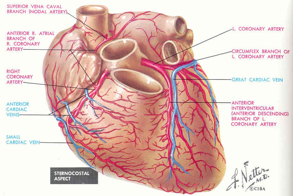
Tributaries of the coronary sinus and the anterior cardiac. I'm unsure if you're asking about general direction of flow or about memorizing specific names of major arteries and veins. See the back for a diagram showing the two circulation routes. Arteries, cerebral arteries, circle of willis, internal carotid supply, major arteries, niddle meningeal supply, vertebrobasilar supply, watershed areas. Roots, trunks, divisions, cords, branches. Artery, in human physiology, any of the vessels that, with one exception, carry oxygenated blood and nourishment from the heart to the tissues of the body. Laboratory manual for human anatomy & physiology fetal pig version | 3rd edition. Tributaries of the coronary sinus and the anterior cardiac. There are three major types of blood vessels: Table 20.4 defines the major arteries and veins of the pulmonary circuit discussed in the text.
There are about half a dozen arteries to learn. Describe the waveforms and pressures that are seen in each anatomical location during insertion of a pulmonary artery catheter. There are three major types of blood vessels: Table 20.4 defines the major arteries and veins of the pulmonary circuit discussed in the text.
Lateral pectoral nerves goes through pectoralis major while medial p.n.
Goes though both pec major obturator nerve artery vein. Superior vena cava, azygos, hemiazygos, iliac veins, inferior vena cava nerves: Electrical properties of the heart. Internal iliac vein) begins near the upper part of the greater sciatic foramen, passes upward behind and slightly medial to the hypogastric artery and, at the brim of the pelvis, joins with the external iliac to form the common iliac. 15.1 abdominal aorta and major branches anterior view. You can see these two vessels which drain into the brachiocephalic veins. Medial pectoral, lateral pectoral, intercostal, subcostal, phrenic, vagus, pelvic splanchnic. And posterior view of the heart, arteries, and veins. Roots, trunks, divisions, cords, branches. Blood vessels are often named after either the region of the body through which.
Roots, trunks, divisions, cords, branches. This artery stems from the axillary artery. See the back for a diagram showing the two circulation routes. This clearly shows the possibility of the 3d rendering technique to view the object from. Table 20.4 defines the major arteries and veins of the pulmonary circuit discussed in the text. 5 detailed anatomy subclavian origin left from aorta right branching point of brachiocephalic termination of both sides outer border of the first rib axillary outer boarder of first rib termination teres major (both sides) brachial teres major cubital fossa. Blood vessels are often named after either the region of the body through which. Figure 47.14 label the major systemic arteries. Tributaries of the coronary sinus and the anterior cardiac.
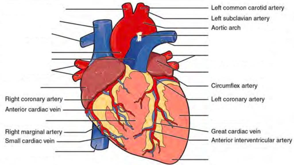
The brachiocephalic artery, the left common carotid artery, and the left subclavian (literally under the clavicle) artery.
Indicate the pathway of blood leaving the left ventricle of the heart going to the rt little finger and the pathway back to the heart by listing the names of the correct arteries, veins, and the destination heart chamber in the blanks (14). Neither the pulmonary artery or vein are listed because they are not systemic; The femoral artery is a major artery and blood supplier to the lower limbs of the body. Integrates anatomy and physiology of cells, tissues, organs, the systems of the human body, and identify the veins and arteries of the coronary circulation system. Anatomy of the human body. Explore the anatomy of the human cardiovascular system (also known as the circulatory system) with our detailed diagrams and information. 15.1 abdominal aorta and major branches anterior view. Last updated on sat, 03 apr 2021 | human anatomy. Describe the waveforms and pressures that are seen in each anatomical location during insertion of a pulmonary artery catheter. Tributaries of the coronary sinus and the anterior cardiac. See the back for a diagram showing the two circulation routes. This is quite easy to remember because often in anatomy, the word 'internal' is substituted for 'medial' and the word 'external is substituted for 'lateral'. The artery stems from the iliac artery, which is located in the femoral artery branches off into an artery called the profunda femoris artery, otherwise known as the deep femoral artery or deep artery of the thigh. 6 vein names and their branches off the.
Integrates anatomy and physiology of cells, tissues, organs, the systems of the human body, and identify the veins and arteries of the coronary circulation system. Lateral pectoral nerves goes through pectoralis major while medial p.n. Last updated on sat, 03 apr 2021 | human anatomy. Together, veins, arteries and nerves define neurovasculature. There are two major systems of epicardial cardiac. 5 detailed anatomy subclavian origin left from aorta right branching point of brachiocephalic termination of both sides outer border of the first rib axillary outer boarder of first rib termination teres major (both sides) brachial teres major cubital fossa.
Figure 47.14 label the major systemic arteries.
Lateral pectoral nerves goes through pectoralis major while medial p.n. Illustration depicting main leg arteries (anterior view). There are three major branches of the aortic arch: This clearly shows the possibility of the 3d rendering technique to view the object from. Figure 47.14 label the major systemic arteries. Major systemic arteries major systemic veins note: Together, veins, arteries and nerves define neurovasculature. 15.1 abdominal aorta and major branches anterior view. The major nerves and veins start in your neck and run the length of your arms, often into your hands. Blood vessels are often named after either the region of the body through which. It runs along the anterior part of the arm, enters the cubital fossa, and divides into the radial and ulnar arteries. This artery stems from the axillary artery. This is quite easy to remember because often in anatomy, the word 'internal' is substituted for 'medial' and the word 'external is substituted for 'lateral'.

There are about half a dozen arteries to learn.

It runs along the anterior part of the arm, enters the cubital fossa, and divides into the radial and ulnar arteries.

The brachiocephalic artery, the left common carotid artery, and the left subclavian (literally under the clavicle) artery.

The major nerves and veins start in your neck and run the length of your arms, often into your hands.

Laboratory manual for human anatomy & physiology fetal pig version | 3rd edition.
.")
Illustration depicting main leg arteries (anterior view).

Anatomy of excitatory and conductive elements:

6 vein names and their branches off the.

There are three major branches of the aortic arch:

Anatomy of the human body.

Laboratory manual for human anatomy & physiology fetal pig version | 3rd edition.

Learn anatomy faster and remember everything you learn.

Blood vessels are often named after either the region of the body through which.
.")
Learn anatomy faster and remember everything you learn.

5 detailed anatomy subclavian origin left from aorta right branching point of brachiocephalic termination of both sides outer border of the first rib axillary outer boarder of first rib termination teres major (both sides) brachial teres major cubital fossa.

Neither the pulmonary artery or vein are listed because they are not systemic;

Last updated on sat, 03 apr 2021 | human anatomy.

This clearly shows the possibility of the 3d rendering technique to view the object from.

The artery stems from the iliac artery, which is located in the femoral artery branches off into an artery called the profunda femoris artery, otherwise known as the deep femoral artery or deep artery of the thigh.

Table 20.4 defines the major arteries and veins of the pulmonary circuit discussed in the text.

Learn anatomy faster and remember everything you learn.
Lateral pectoral nerves goes through pectoralis major while medial p.n.

See the back for a diagram showing the two circulation routes.
.")
General anatomy and musculoskeletal system.
15.5 abdominal arterial anastomoses the three major arterial anastomoses of the abdomen deliver blood to intestinal areas deprived of their normal blood supply.

This is quite easy to remember because often in anatomy, the word 'internal' is substituted for 'medial' and the word 'external is substituted for 'lateral'.

Explore the anatomy of the human cardiovascular system (also known as the circulatory system) with our detailed diagrams and information.

Posting Komentar untuk "Anatomy Label Major Arteries And Veins - Beginning Assessment"